Vision

Introduction
The human visual system is a complex and sophisticated network that allows us to perceive and interpret the world around us. It involves not just the eyes but also numerous components within the brain that process visual information. Understanding how each element functions provides insight into how we see, interpret depth, color, movement, and how these processes can be mimicked in artificial vision systems. This documentation will break down the human visual system into its various components, provide definitions, explain how images are processed, and compare these processes to those in AI/robot vision systems.
Glossary of Components and Structures in the Human Visual System

1. Eye Anatomy

Cornea: The transparent, dome-shaped surface that covers the front of the eye. It helps to focus incoming light.
Pupil: The adjustable opening in the center of the iris that controls the amount of light entering the eye.
Iris: The colored part of the eye surrounding the pupil that adjusts the pupil size.
Lens: A transparent structure behind the pupil that changes shape to focus light on the retina.
Retina: The light-sensitive layer at the back of the eye containing photoreceptor cells (rods and cones) that convert light into neural signals.
Rods: Photoreceptor cells sensitive to low light levels, crucial for night vision and peripheral vision.
Cones: Photoreceptor cells responsible for color vision and high spatial acuity.
Ganglion Cells: Neurons located in the retina that receive input from rods and cones via bipolar cells and transmit visual information to the brain via the optic nerve.
Optic Nerve: The bundle of nerve fibers that transmits visual information from the retina to the brain.
Macula: The central area of the retina responsible for high-resolution vision.
Fovea: A small pit in the macula densely packed with cones, providing the sharpest vision.
2. Brain Structures Involved in Vision
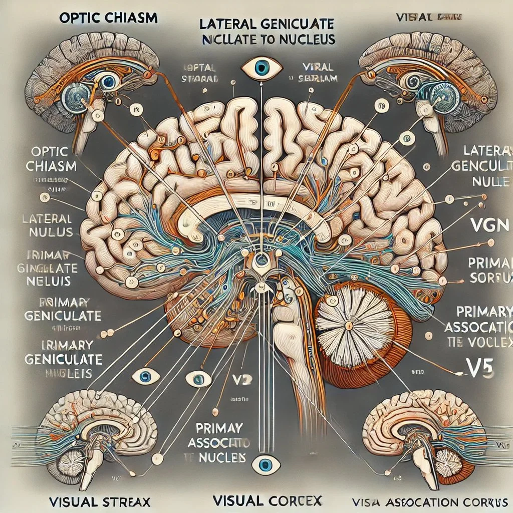
Optic Chiasm: The X-shaped structure where the optic nerves partially cross, allowing visual information from each eye to be processed by both hemispheres of the brain.
Lateral Geniculate Nucleus (LGN): A relay center in the thalamus that processes visual information before it reaches the visual cortex.
Primary Visual Cortex (V1): The first stage of cortical processing of visual information, located in the occipital lobe.
Visual Association Areas (V2-V5): Regions involved in higher-order processing such as color (V4) and motion (V5/MT).
Dorsal Stream ("Where Pathway"): Processes spatial awareness and movement.
Ventral Stream ("What Pathway"): Processes object recognition and form representation.

3. Visual Processing Concepts
Segmentation: The process of dividing the visual scene into distinct objects and background.
Background and Foreground: Elements in the visual field that are perceived as distant (background) or closer (foreground).
Horizon Line: The apparent line that separates the earth from the sky, crucial for spatial orientation.
Vertical Line: Lines that run up and down in the visual field, important for perceiving depth and structure.
Binocular Convergence: The inward movement of both eyes toward each other to maintain single binocular vision when viewing an object.
Focal Point: The point at which the eyes are aimed to focus on an object.
Focal Plane: The plane where objects are in sharp focus on the retina.
Binocular Rivalry: A phenomenon where perception alternates between different images presented to each eye.
Binocular Fusion: The process by which the brain combines the two slightly different images from each eye into a single image.

Detailed Breakdown of the Human Vision System
1. Light Entry and Initial Focusing
Cornea and Aqueous Humor: Light first enters the eye through the cornea, which, along with the aqueous humor (fluid between the cornea and lens), begins the focusing process due to its refractive properties.
Pupil and Iris: The iris adjusts the size of the pupil to control the amount of light entering the eye. In bright conditions, the pupil constricts; in dim conditions, it dilates.
Lens Accommodation: The lens adjusts its shape (accommodation) to fine-tune the focus of light onto the retina. This is achieved by the ciliary muscles changing the lens's curvature.
2. Phototransduction in the Retina
Retina Structure: The retina contains layers of neurons, including photoreceptors (rods and cones), bipolar cells, and ganglion cells.
Rods and Cones:
Rods: Approximately 120 million rods are sensitive to light intensity but not color. They function well in low-light conditions and are primarily located in the peripheral retina.
Cones: Around 6 million cones detect color and are concentrated in the fovea. There are three types of cones sensitive to red, green, and blue light wavelengths.
Phototransduction Process: When photons hit photoreceptors, they trigger chemical changes in photopigments, leading to a change in the electrical potential of the cell and the generation of neural signals.
3. Signal Transmission to the Brain
Bipolar Cells: Receive input from rods and cones and transmit signals to ganglion cells.
Ganglion Cells: Their axons form the optic nerve, transmitting visual information to the brain.
Optic Nerve and Optic Chiasm: The optic nerves from both eyes meet at the optic chiasm, where fibers partially cross over to allow visual information from the right visual field to be processed by the left hemisphere and vice versa.
4. Visual Processing in the Brain
Lateral Geniculate Nucleus (LGN): Acts as a relay and processing station, refining visual signals before they reach the cortex.
Primary Visual Cortex (V1): Processes basic visual components such as edges, orientations, and motion.
Visual Association Areas:
V2: Further processes features like orientation, spatial frequency, and color.
V3: Involved in processing motion and depth.
V4: Specialized in color perception and form recognition.
V5/MT: Critical for motion detection.
Dorsal and Ventral Streams:
Dorsal Stream: Handles spatial awareness, guiding actions, and recognizing where objects are in space.
Ventral Stream: Involved in object recognition, identifying what objects are.
5. Image Processing Mechanisms
Segmentation: The visual system separates the visual scene into individual objects and background through edge detection, texture differences, and motion cues.
Depth Perception:
Binocular Cues: Using the slight difference between images in each eye (binocular disparity) to perceive depth.
Convergence: The degree to which eyes turn inward to focus on an object helps determine its distance.
Motion Detection: Specialized cells detect movement direction and speed.
Color Processing: Cones respond to different wavelengths, and the brain combines these signals to produce the perception of color.
Binocular Rivalry and Fusion:
Rivalry: Occurs when each eye is presented with significantly different images; perception alternates between them.
Fusion: The normal process of combining similar images from each eye into a single perception.
Visual Processing Concepts Explained
1. Segmentation
The brain's ability to distinguish different objects within a visual scene by detecting edges, contrasts, and discontinuities.
2. Background and Foreground Processing
Differentiating objects (foreground) from their surroundings (background) using cues like size, motion, and focus.
3. Horizon Line and Vertical Lines
Horizon Line: Helps in perceiving spatial orientation and depth; the brain uses it to interpret the position of objects in space.
Vertical Lines: Assist in recognizing structures and spatial relationships, crucial for balance and navigation.
4. Dual Eye Convergence Points and Focal Plane
Convergence Points: The point where the eyes' lines of sight meet; important for depth perception.
Focal Plane: The plane at which objects are in sharp focus on the retina, adjusted by lens accommodation.
Far and Near Cells: Cells in the visual cortex that respond preferentially to objects at different distances.
5. Binocular Rivalry and Fusion
The interplay between the two eyes' inputs, where similar images are fused for depth perception, and dissimilar images may cause rivalry, leading to alternating perception.